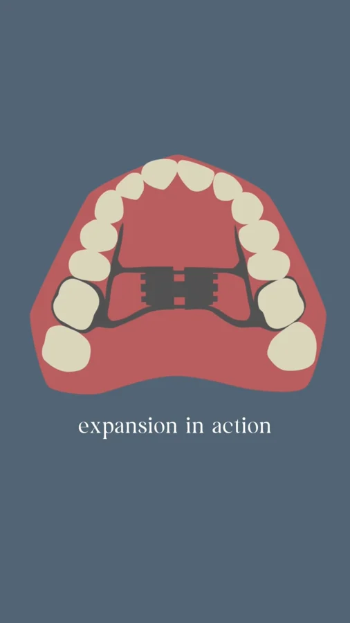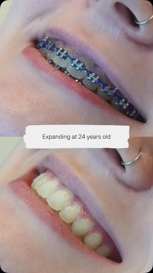

Two spaces with a structural connection
The published caption uses a memorable explanation: the roof of the mouth and the floor of the nose share a structural relationship. The upper jaw is not an isolated platform that holds teeth. It contributes to the framework below the nasal cavity, which helps explain why a treatment that changes upper-jaw width may also influence nasal space.
For a patient, this makes airway-focused assessment easier to understand. The clinician is considering neighboring structures together. Looking at the teeth, palate, and nose as connected parts can reveal questions that a conversation about straight teeth alone might never bring to the surface.
What expansion research has measured
In a 2022 randomized trial comparing three expander designs, patients with narrow upper jaws showed increases in measured nasal cross-sectional area and nasal volume. A separate small imaging study in children with sleep apnea and malocclusion also documented changes in nasal width and upper-airway volumes after expansion.
These findings support the anatomical connection described in the post. They do not tell us exactly how much change a particular patient will experience. Appliance comparisons and small before-and-after studies answer specific questions; the clinical examination still needs to establish why expansion is appropriate and what success should mean for that individual.
A wider space is one part of the assessment
The useful next question is where a patient's difficulty is coming from. A narrow palate may be relevant, but a breathing complaint cannot be fully understood from the appearance of the dental arch. The care team needs to connect the structural finding with the person's history and the situations in which breathing feels difficult.
This is particularly important when symptoms occur during sleep. A drawing of the palate or an image showing more space can explain anatomy, but it cannot show the complete pattern of breathing through a night. Structural evaluation and appropriate medical assessment contribute different pieces of information to a coordinated plan.
Ask to see the connection in your own records
During a consultation, ask the clinician to show you the specific upper-jaw concern and explain how it relates to the proposed treatment. Useful questions include what will be measured, which changes are expected in the bite, and how any breathing-related goals will be assessed separately.
There is no need to arrive with a diagnosis or to decide in advance that an expander is the answer. Start with what you have noticed. The purpose of understanding this anatomy is to ask better questions and make a more informed decision about care, with a plan that reflects your own findings rather than a general illustration.
The shared nasomaxillary anatomy explains a mechanism, not a guaranteed result
The hard palate and nasal floor are anatomically related components of the nasomaxillary complex. A transverse orthopedic change in this region therefore has a plausible relationship to nasal dimensions. That relationship is more specific than saying the mouth and airway are simply “connected”: it identifies an anatomic structure that may change and a measurable consequence that can be investigated.
However, the nasal airway also includes soft tissues and regions that are not represented by the width of a dental arch. A clinician must consider where resistance or obstruction is suspected and whether the documented transverse deficiency is relevant to that concern. The appearance of a high palate is a reason for assessment, not a numerical estimate of nasal resistance. The anatomical hypothesis and the patient’s actual functional problem must be evaluated together.
What a paired dental and respiratory outcome record should show
An informative record separates structural correction from respiratory follow-up. Dental documentation may describe arch coordination and the change in a transverse discrepancy. Respiratory documentation should specify the symptom or measurement being followed and the responsible medical clinician. Recording both avoids allowing one successful endpoint to stand in for another that was never measured.
This approach is especially important when a patient reports better breathing after orthodontic treatment. The report is clinically relevant, but the interpretation depends on baseline symptoms, concurrent nasal treatment, and the follow-up interval. If OSA was previously diagnosed, it remains a medical condition requiring appropriate reassessment even when daytime nasal breathing feels easier. The shared anatomy gives the care team a useful reason to coordinate; it does not remove the need to distinguish nasal patency from sleep-state pharyngeal obstruction.
Bring the conversation to your visit.
- Can you show me the upper-jaw finding that matters in my case?
- Which expected changes involve the bite, and which involve nasal function?
- How will any persistent breathing symptoms be assessed after treatment?
Supporting studies & resources
These resources add clinical context to the original post. Each publication should be read with its study population, methods, and outcomes in mind.
- Evaluation of the effects of different rapid maxillary expansion appliances on airway by acoustic rhinometry: A randomized clinical trial Gokce et al. · International Journal of Pediatric Otorhinolaryngology · 2022 · Randomized trial
- Cone beam CT evaluation of skeletal and nasomaxillary complex volume changes after rapid maxillary expansion in OSA children Pirelli et al. · Sleep Medicine · 2021 · Uncontrolled before-and-after study
- Policy on Obstructive Sleep Apnea (OSA) American Academy of Pediatric Dentistry · 2026 · Professional policy
- Polygraphic evaluation of the effects of different rapid maxillary expansion appliances on sleep quality: A randomized clinical trial Gokce et al. · Sleep and Breathing · 2023 · Randomized trial