
Airway-focused orthodontics
The way teeth fit is part of a bigger picture. We consider the upper jaw, lower jaw, tongue space, and developing bite alongside the breathing and sleep concerns you bring to your visit.
ExploreExplore the two clinical sequences from our visual guide. The images illustrate dental and structural changes; they do not establish a particular breathing or sleep result.
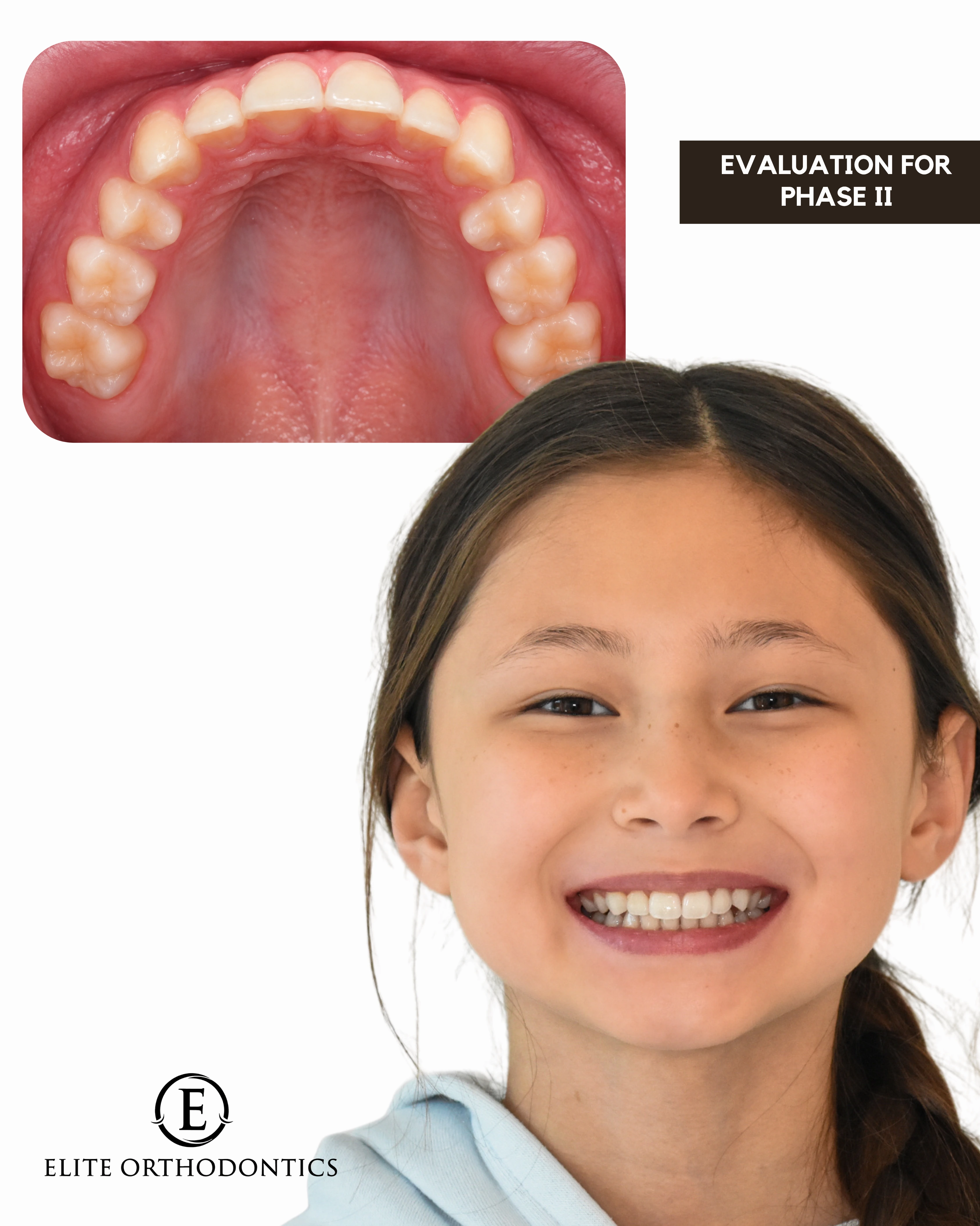
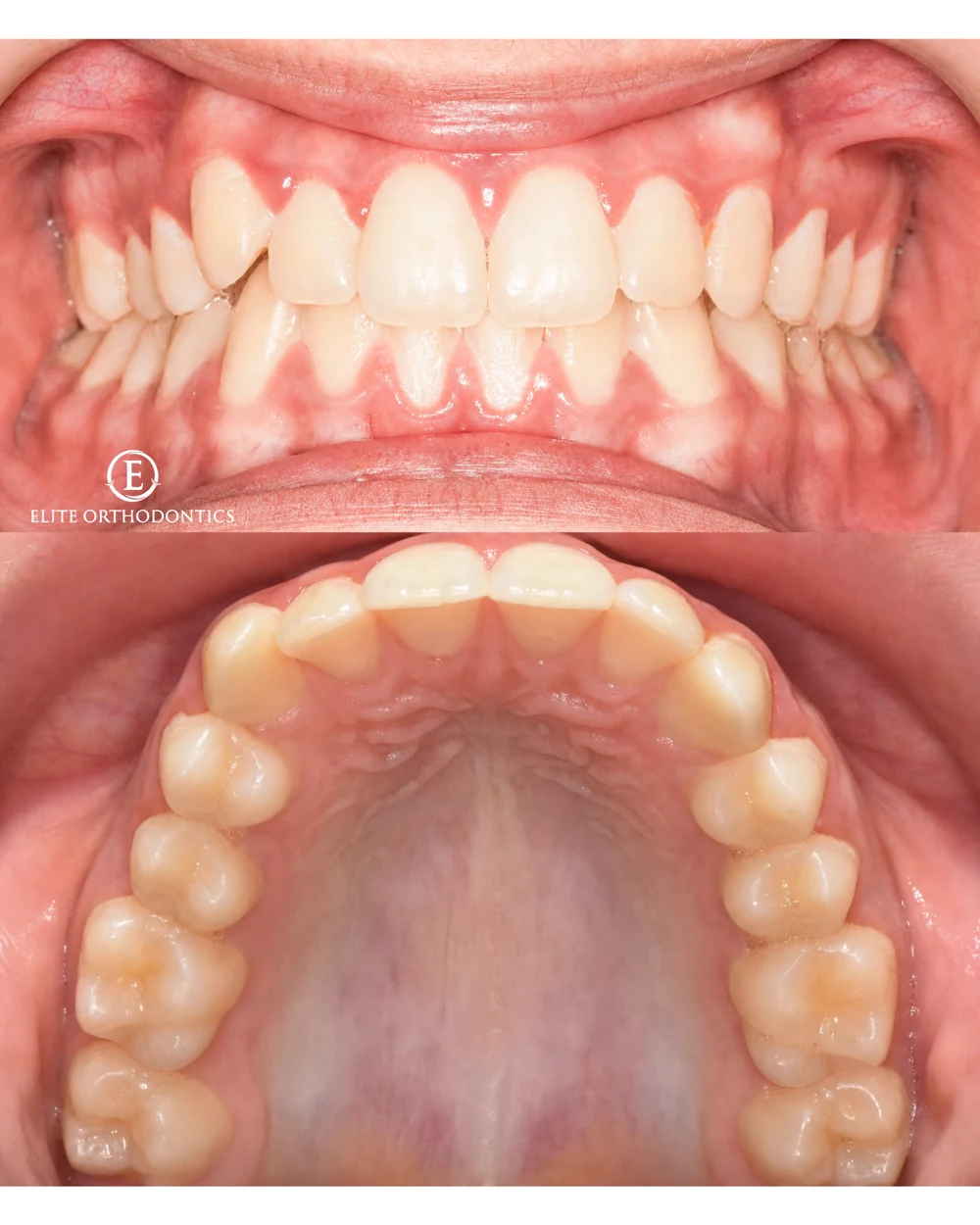
Eight images follow the supplied sequence of crowding and palatal expansion. Use the arrows, keyboard, or swipe to explore.
An individual clinical example. Dental changes do not establish a breathing or sleep outcome. Results and treatment needs vary.
Explore the clinical gallery
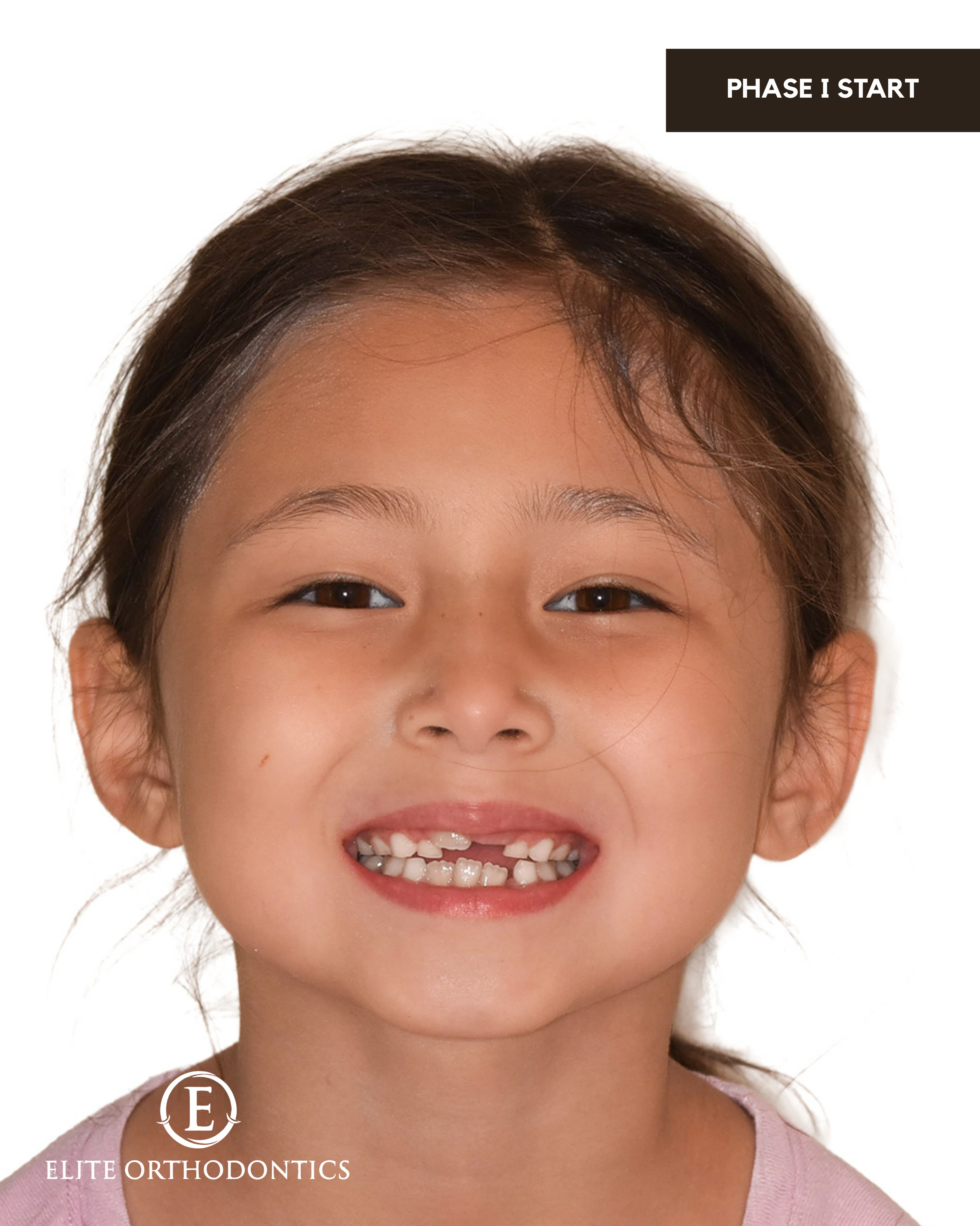
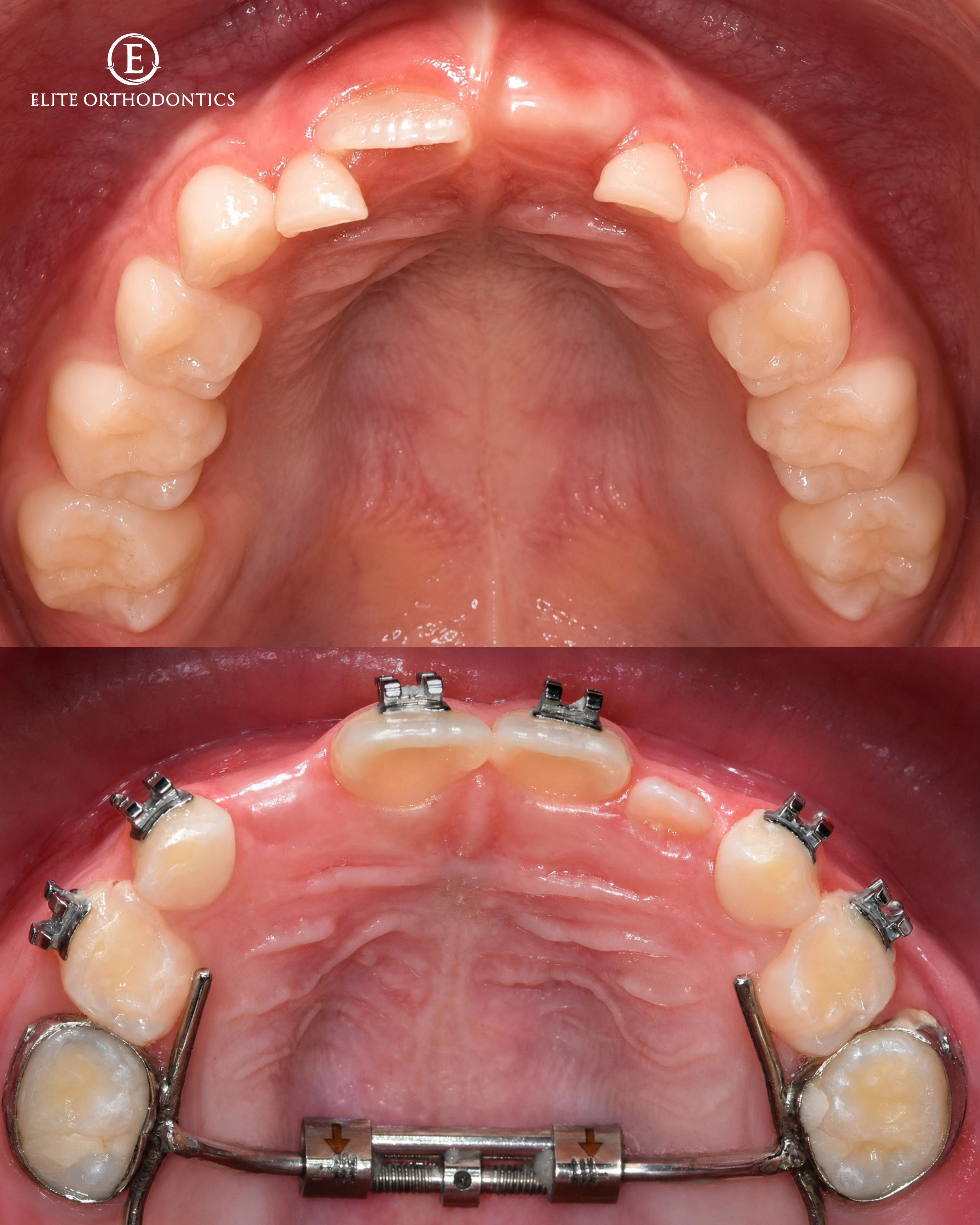
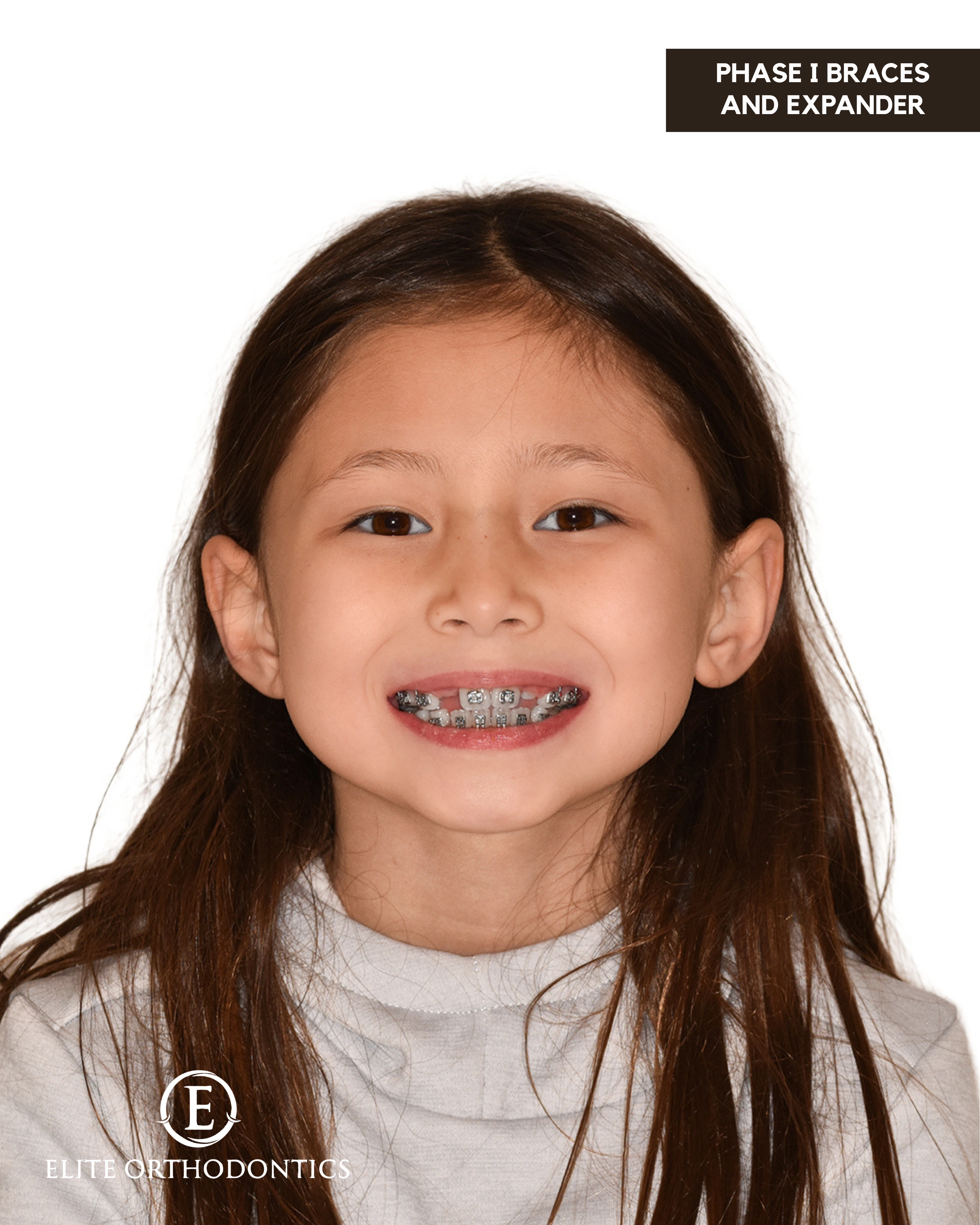

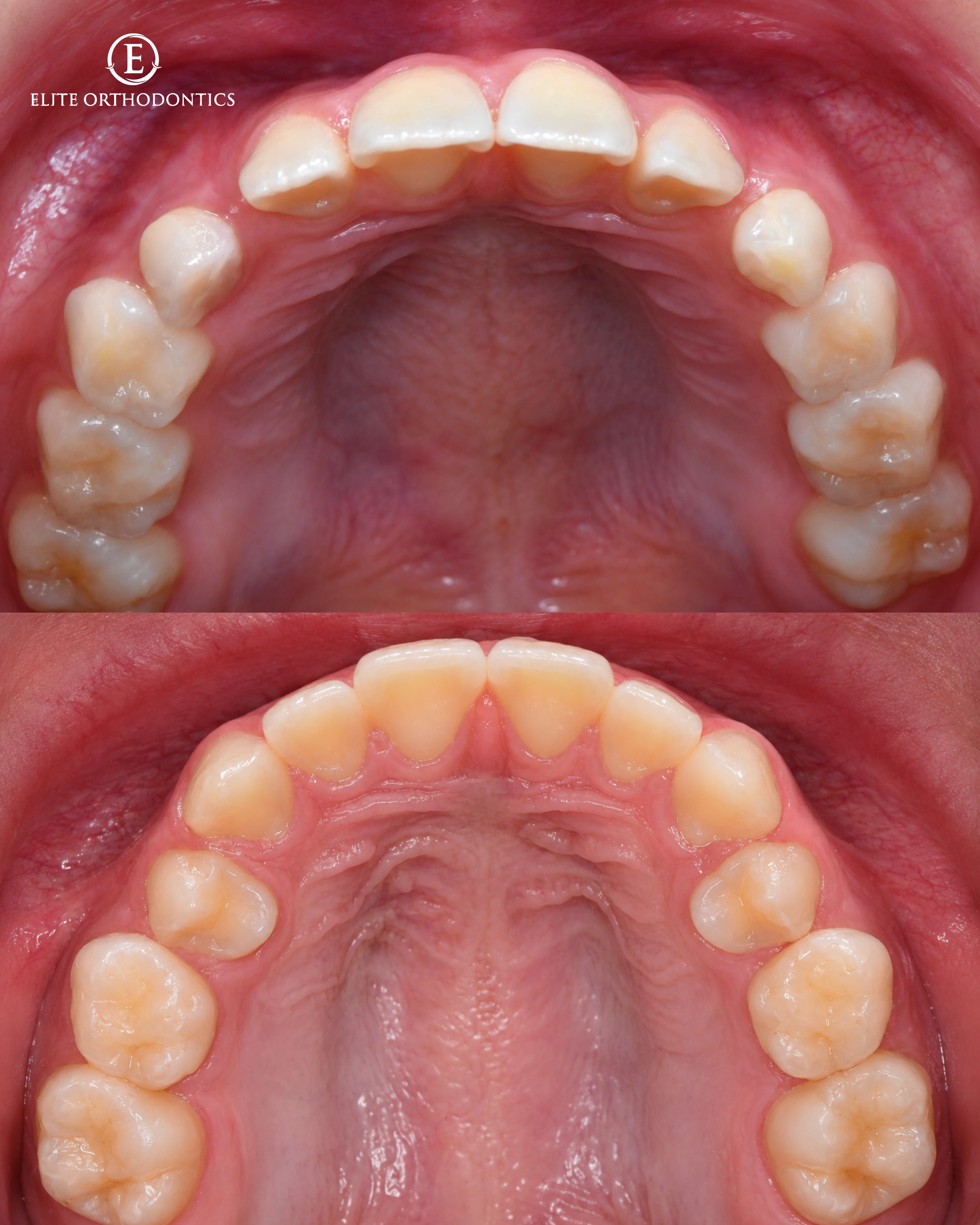

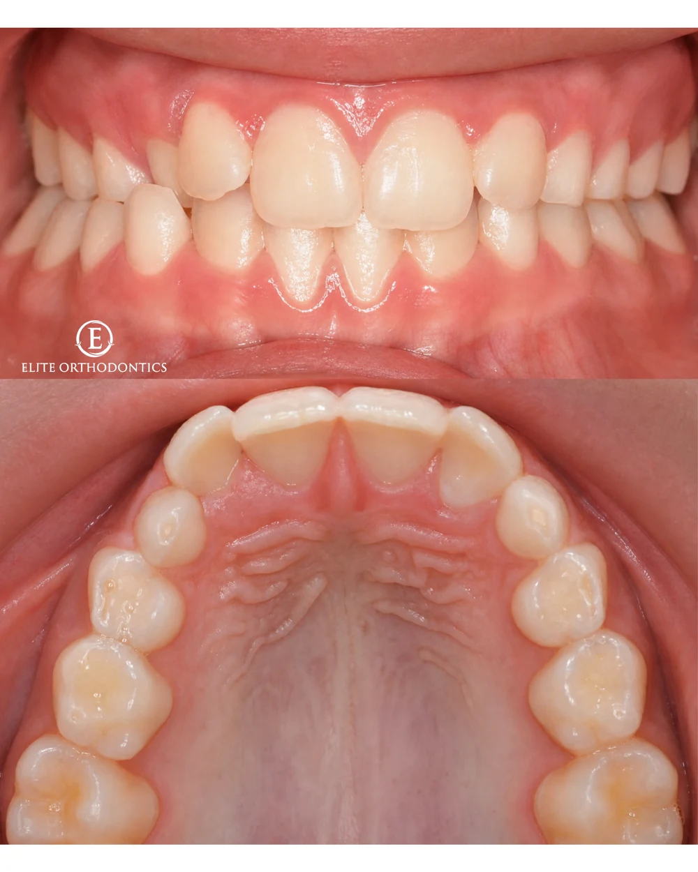
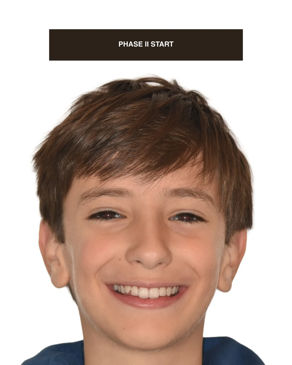
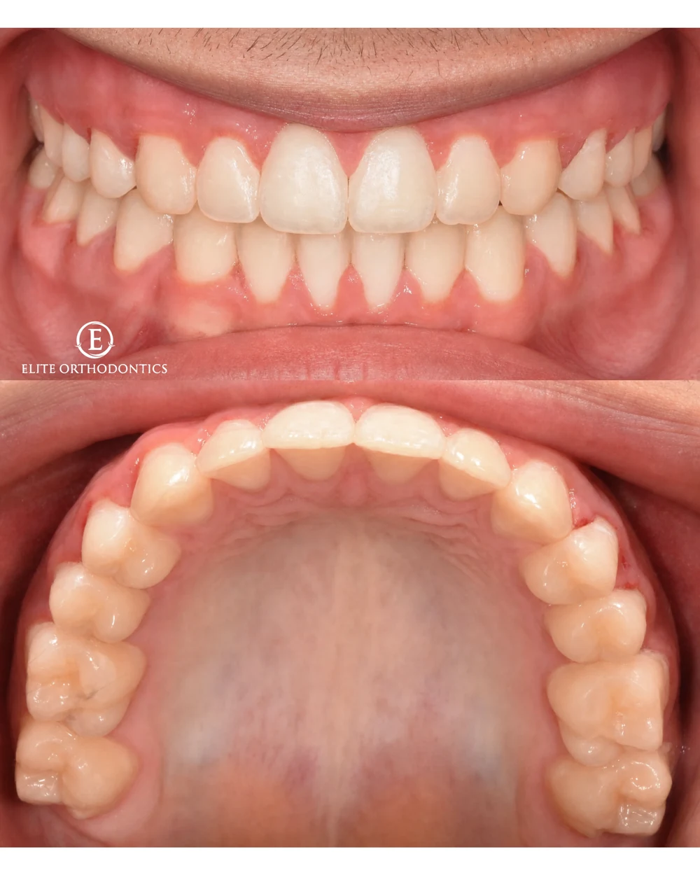
Six images follow the supplied sequence of crossbite and upper-arch development. Every treatment plan and result is individual.
An individual clinical example. Dental changes do not establish a breathing or sleep outcome. Results and treatment needs vary.
Explore the clinical gallery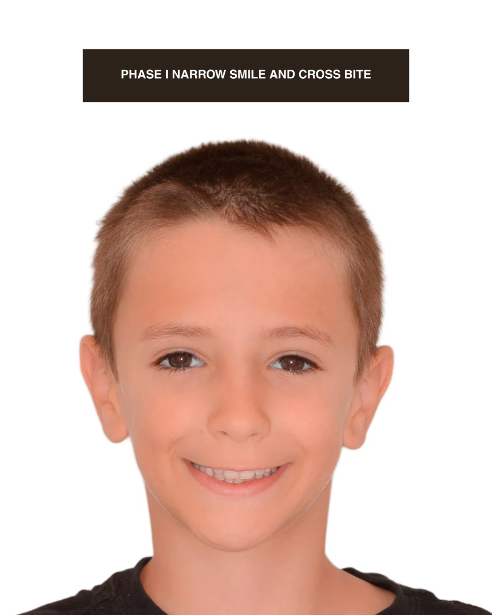

The way teeth fit is part of a bigger picture. We consider the upper jaw, lower jaw, tongue space, and developing bite alongside the breathing and sleep concerns you bring to your visit.
ExploreThe palate is also the floor of the nose. When the upper jaw is constricted, expansion can address a structural limitation that matters to the bite and, in selected patients, the nasal airway.
ExploreThe supplied Phase I and crossbite galleries allow the reader to observe the dental relationships documented in the images. Relevant observations include the transverse relationship of the upper and lower arches, available eruption space, tooth alignment, and intercuspation. Image angle, magnification, and head position can affect apparent change, so precise measurements should come from appropriately standardized clinical records. A visual comparison is useful for education, but it is not equivalent to a complete diagnostic work-up or a quantified treatment-outcome study.
A posterior crossbite can involve dental inclination, skeletal transverse discrepancy, or a functional mandibular shift. Crowding reflects the relationship between tooth dimensions and available arch space and does not, by itself, identify the cause. The photographs should therefore prompt diagnostic questions: Was a functional shift present? Which component of the discrepancy was treated? What was the dentition stage? Those questions cannot all be answered by appearance alone and should not be filled in with assumptions about an individual case.
A clinically interpretable case report would include the presenting concern, medical and dental history, examination findings, diagnosis, rationale for treatment selection, relevant baseline records, treatment sequence, and follow-up. It would state the outcome measures and any adverse events or additional care. When respiratory improvement is a treatment objective, the report should identify how that outcome was assessed. The supplied image galleries do not contain a complete set of these records, so this site does not assign unreported sleep-study changes, symptom scores, or long-term results to the patients shown.
An immediate change in alignment or arch form answers a different question from stability after retention. Follow-up may examine occlusal contacts, eruption, periodontal condition, appliance effects, and the persistence of the original functional concern. A respiratory diagnosis, if present, requires follow-up within the appropriate medical pathway. This separation allows a case to demonstrate a dental result without being presented as proof of a sleep outcome. It also makes the educational value of the images more precise and useful to referring clinicians.
Supporting sources: Gokce et al.: Nasal airway dimensions after expansion, 2022 AAPD: Policy on Obstructive Sleep Apnea, 2026
Bring your breathing, sleep, and development concerns into one conversation. Begin with our online airway evaluation.
Start airway evaluation (opens in a new tab)Educational screening only. An in-person assessment determines diagnosis and treatment options.